

We’re into the final stretch of 2026 already, and the extracellular vesicle (EV) community has been busy!
Here’s a roundup of selected highlights published so far this year that used Izon’s qEV size exclusion chromatography (SEC) columns, the Automatic Fraction Collector (AFC) and the Exoid. For each paper, we look at what the team found and how Izon’s tools fitted into their workflows.
Small and Large Extracellular Vesicles in Circulation of Diffuse Large B-Cell Lymphoma Patients Originate from Different Cell Types of the Tumor Microenvironment
Blood tests that read EVs, known as liquid biopsies, could one day help track cancer without a tissue sample. But which EVs should those tests look at? Maltoni et al. asked whether small and large EVs in the blood of patients with diffuse large B-cell lymphoma come from different cells.¹ The team ran plasma and cell culture medium through qEVoriginal 70 nm columns on the AFC to collect small EVs. They pelleted large EVs by ultracentrifugation and sized both with the Exoid.
The answer was yes! Small EVs came mainly from the cancer cells, while large EVs came more from immune cells around the tumour, such as macrophages, T cells and natural killer cells. The authors say this has “important implications for the development of EV-based liquid biopsy strategies”, since a test may need to target a particular EV size depending on which cells it aims to track. They also point to SEC’s practical advantages over ultracentrifugation, including “reduced labour intensity”, noting that ultracentrifugation “is generally not considered scalable for clinical practice”.
Workflow for the Isolation and Characterisation of Human Milk Extracellular Vesicles (HMEVs) and Their Inflammatory Biomarker Profile
Human milk EVs carry oxylipins, fat-derived signalling molecules that help calm inflammation and may be relevant to necrotising enterocolitis, a serious gut disease in preterm babies. But milk is a tricky sample, full of fat and casein. Moreno-Casillas et al. set out to build a better workflow.² They used qEV 35 nm columns on the AFC, followed by ultracentrifugation, and measured the EVs with the Exoid.
Compared with ultracentrifugation alone, the qEV-based workflow gave about three times the vesicle concentration, while needing only 7 mL of milk instead of 25 mL. The authors found it “superior… in terms of vesicle recovery, purity and structural integrity”, and chose SEC because, as they note, it “stands out for its simplicity, scalability and its ability to preserve vesicle integrity”. Using milk from 10 donors, they then profiled the oxylipins inside the EVs and detected arachidonic acid-derived EETs in human milk EVs for the first time. The team describes the result as a “reproducible, high-yield workflow adaptable to both small- and large-scale research settings”.
Venom vesicles from the parasitoid Ganaspis hookeri facilitate venom protein entry into host immune cells
Parasitoid wasps lay their eggs inside fruit fly larvae, then have to get past the host’s immune defences. Bretz et al. found that Ganaspis hookeri packs its venom proteins into extracellular vesicles (venom EVs) that deliver them into the host’s immune cells (plasmatocytes).³
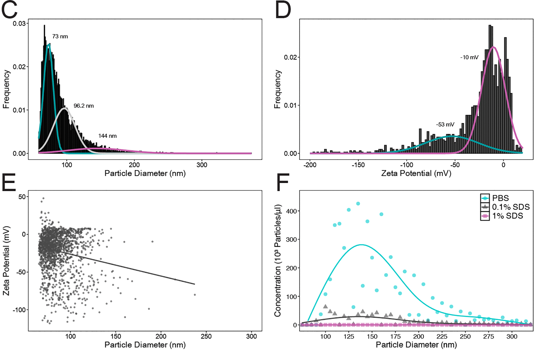
To characterise these venom vesicles, the team purified the vesicles on a sucrose gradient and used the Exoid with an NP100 nanopore. Because tunable resistive pulse sensing (TRPS) measures particles one at a time, it gave them the size and zeta potential of each vesicle, not just an average. The data revealed three size populations (around 73, 96 and 145 nm) and two distinct surface charge groups (around −10 and −53 mV). When they treated the venom with detergent, vesicle numbers dropped sharply and almost none were left at the highest dose. This supports the conclusion that the particles are membrane-bound vesicles (Figure 1). The authors propose a model in which venom proteins are packaged into vesicles and carried into host cells to suppress immunity.
RAB27A silencing modulates miR-30d-5p expression and extracellular vesicle non-coding RNA cargo in diabetic podocytes
Diabetic kidney disease is one of the leading causes of kidney failure, and one of its earliest signs is damage to podocytes, the cells that help filter blood. But what messages do these stressed cells send out in their EVs? Martinez-Arroyo et al. grew human podocytes in high glucose to mimic diabetes, and switched off RAB27A, a gene that controls EV release.⁴ The team isolated EVs from the cell culture medium with qEVoriginal columns and measured them on the Exoid.
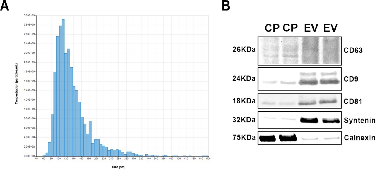
On the Exoid, the EVs peaked at around 90–120 nm, and they carried the classic EV markers CD63, CD9, CD81 and syntenin (Figure 2). Both high glucose and RAB27A silencing changed the EVs’ RNA cargo, and miR-30d-5p was the only microRNA consistently affected. It targets SMAD1, which is involved in antifibrotic signalling. The authors suggest miR-30d-5p could be a mediator of these pathways under glucose stress.
Comparative Profiling of Mouse and Human Microglial Small Extracellular Vesicles Reveals Conserved Core Functions with Distinct miRNA Signatures
Much EV research in neuroscience starts in mice, but how well do the findings carry over to humans? Bayat et al. compared small EVs from mouse and human microglia, the brain’s immune cells.⁵ They isolated EVs from cell culture medium with qEV2 35 nm columns, using the AFC to collect equal-volume fractions.
Checking each fraction showed exactly where the EVs ended and contamination began. Fractions 2–5 were rich in the EV marker TSG101 and free of calnexin, a contamination marker, so the team pooled only these. Using only these clean fractions, the team found that EVs from both species had similar core effects on human Schwann cells. However, human EVs protected better against oxidative stress and although the two species shared most of their microRNAs, each had its own distinct signature.
Biomechanical strain alters the microRNA cargo of primary myogenic cell-derived extracellular vesicles associated with a differentiation phenotype
Every time a muscle moves, its cells are pulled and stretched. Does that change the messages they send? Williams et al. stretched mouse muscle precursor cells, then isolated their EVs using qEVoriginal 35 nm columns and AFC, collecting ten 0.5 mL fractions.⁶ They pooled the clean “EV-rich, protein-poor” fractions for small RNA sequencing and for treating new muscle cells.
High-strain stretching raised EV output 2.5-fold, while long, low-level strain raised miR-222 in the EVs and cells that received these EVs showed changes linked to muscle cell maturation, including more myosin heavy chain. The authors call the work “hypothesis-generating”, but it hints that stretching could one day be used to tune EV cargo for muscle repair.
Ready to isolate EVs for your next study?
- Find the right column in the qEV range.
- See how the Automatic Fraction Collector takes the manual work out of collecting fractions.
- Measure your EVs’ size, concentration and charge with the Exoid and its nanopores.
- Learn more about EV isolation and particle measurement with TRPS.