PARTICLE MEASUREMENT
Extracellular vesicle characterisation you can count on
Measure EV size, concentration and zeta potential at the single-particle level with Izon’s particle measurement systems.
Measurement down to 30 nm
Get a complete picture of your sample, including its smallest EVs.
High-resolution, repeatable sizing
Separate subpopulations into distinct, well-defined peaks, run after run.
Single-particle zeta potential
Characterise EV surface charge alongside size and concentration.
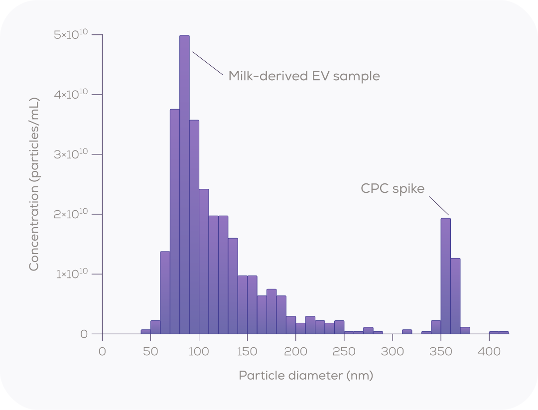
Distinct populations, clearly separated
Single-particle measurement keeps distinct populations distinct. Spike calibration particles into a milk-derived EV sample and the Exoid shows them as their own peak, cleanly separated from the vesicles. You see subpopulations and contaminants, instead of one blended mean.
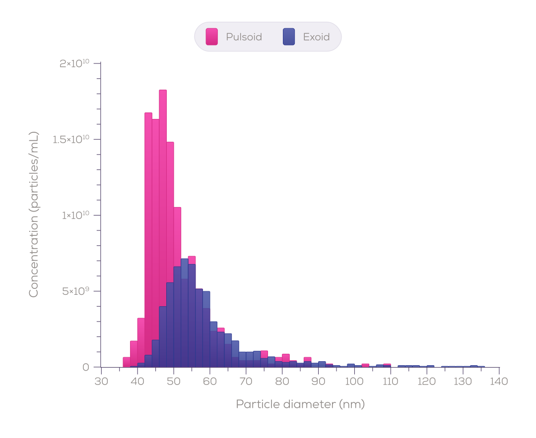
Small EVs, sized and counted
Light scattering weakens steeply as particles get smaller, so small, low-contrast EVs are easy to lose in background noise. Resistive pulse sensing detects each vesicle electrically as it passes through a nanopore on the Pulsoid, so the smallest EVs in your sample are sized and counted.
Resolve what light scattering blurs together
In a published comparison, one four-population bead mixture was measured by tunable resistive pulse sensing (TRPS), NTA and multi-angle DLS. TRPS resolved all four sizes. NTA returned one broad envelope, and MADLS a single mode¹.
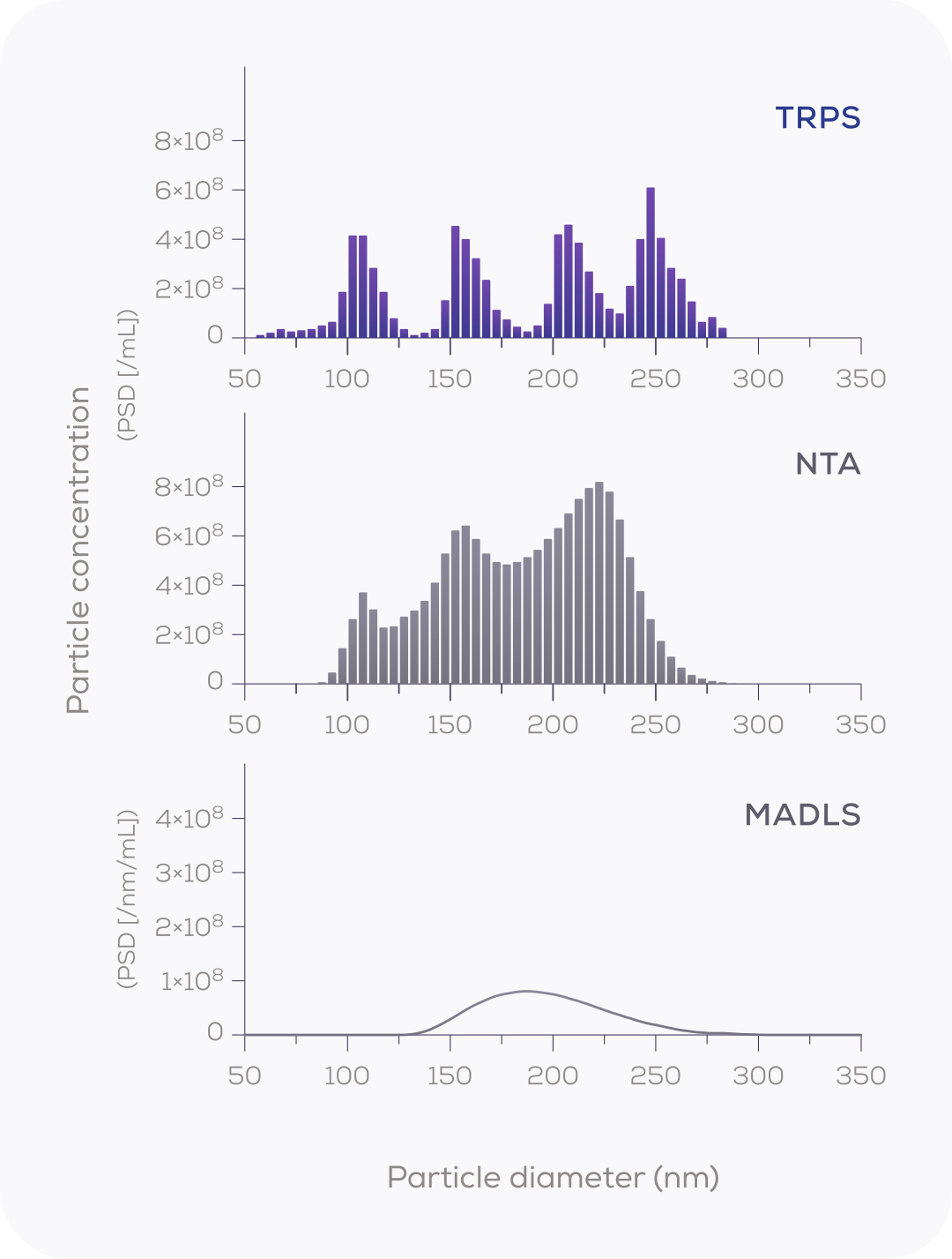
Nanopore sensing vs light-based sizing
Size resolution: the ability to separate particle populations of similar size.
¹ Vogel R. et al. (2021) J Extracell Vesicles 10(3):e12052, Fig. 3. Reproduced under CC BY 4.0. Measured by TRPS.
Choose the right tool for your workflow
Characterise your isolates particle by particle for fundamental research, biomarker discovery, health and disease comparisons, or storage studies. Choose the instrument that fits your samples.
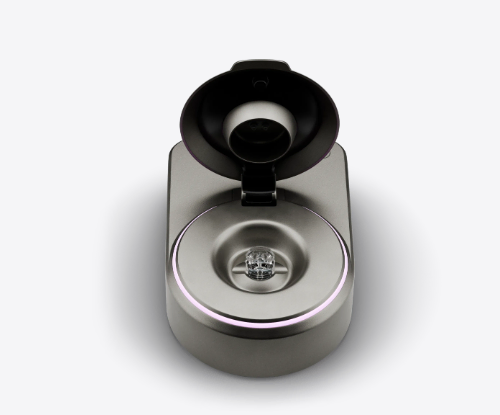
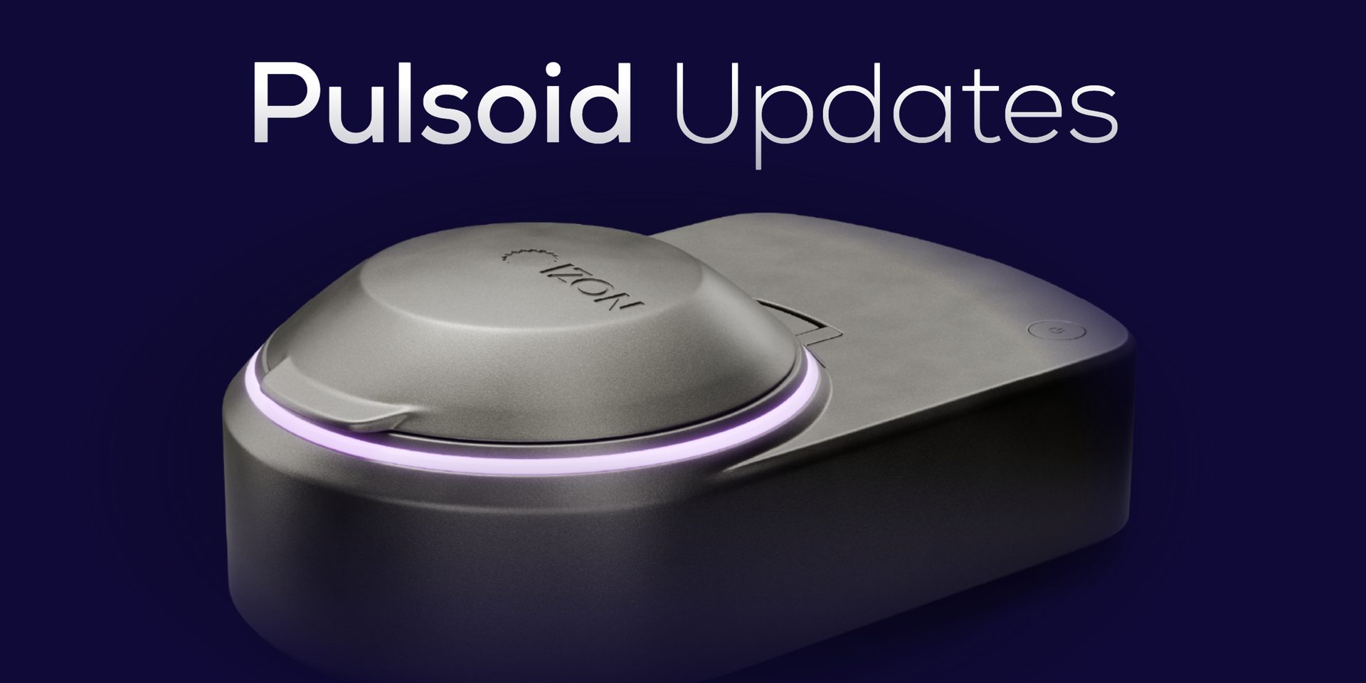
The Pulsoid