Synopsis
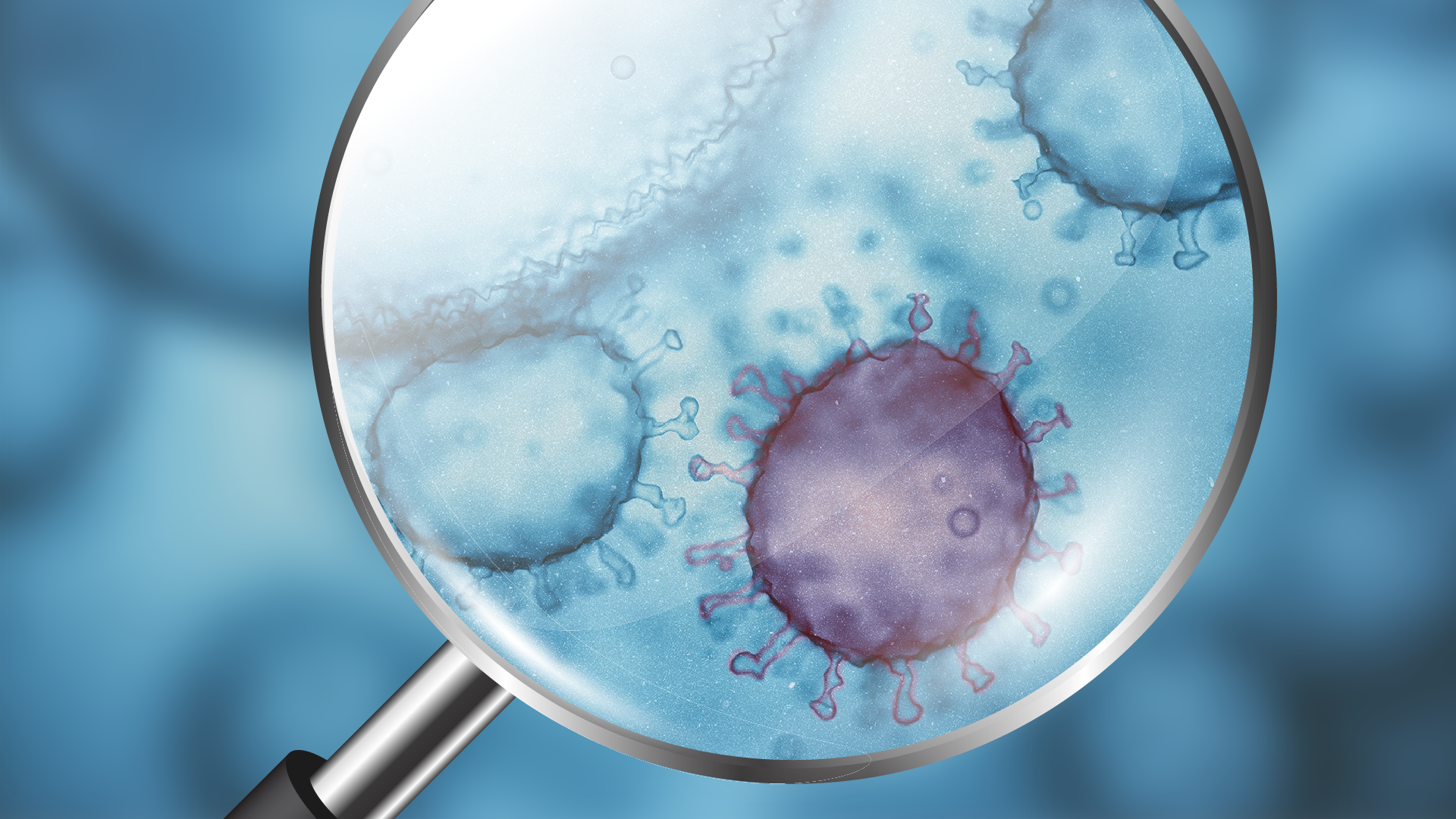
Various viral infections still pose a great threat to human and animal health. A wide range of viral species are surrounded by a lipid shell, the envelope, containing viral and cellular proteins.
The group of enveloped viruses contains species of medical and biotechnical importance such as members of the families Retro- (HIV), Filo- (Ebola), Flavi- (West Nile, Zika, Dengue) and Orthomyxoviridae (Influenza) to name but a few. The contribution of protein elements of the envelope to viral reproduction and pathology is widely studied. We are more interested in the contribution of the lipid elements especially in the behavior of viral (lipid) envelopes after biophysical and biochemical stimuli. Furthermore we try and use this information in the modification of enveloped virus surfaces (i.e. Retro/Lentiviruses) for applications in gene therapy and vaccine development. One important tool we try to develop is multi-parametric single particle detection systems. The latter point is most important for acquiring reliable data on the homogeneity of the samples, answering questions such as: how strongly single viral particles differ from each other regarding morphological, biophysical or biochemical properties? Are different populations recognizable (i.e. contamination by other vesicles/particles or subpopulation of virus particles)? – TRPS provides an in-road into the topic by measuring size and charge of individual particles – key parameters for describing viral populations. Additionally, Since EVs often contaminate viral preparations, for our purposes a negative selection for EV would be most important (i.e. to remove EV from viral samples). However, a quick and reliable, cost-effective purification of virus as provided by qEV columns is highly welcome for our research.
1. How important is single particle measurement to your work?
Very. Somehow comparable to quality control in manufacturing processes: cells produce a large number of particles (not exclusively viral) and in order to do research on them we need to know how closely (or not) they resemble one another.
2. What advantages does the qEV method of virus isolation have for you compared to the other options?
Mostly speed and the simple (and gentle) procedure reducing time and chemical stress. Both means we can keep the particles functional (and/or infectious) as much as possible.

3. How widely understood is this issue of the exosome contamination in the virus sample, given that they are of similar size?
I’d be tempted to say not a lot. As it’s emerging, the interplay between different particle types seems to be pretty close. Mostly the nature of both, the virus and the infected cell will decide. However, drawing clear lines can be very difficult.
4. Can you distinguish the viruses from the background exosomes by innate charge measurement, or perhaps by attaching a charged probe with your virus painting technique?
For the first part, in some cases, I’m guessing yes. However, to my knowledge this has not been tried yet. Choosing the right pair would help. For the second part, not with molecular painting (currently, it doesn’t discriminate between different membrane types. However you may use it to discriminate membrane bound from protein capsid only particles). In any case, the quality of the marker(s) would be the critical issue.
5. Can you use real-time measurement of the dynamic changes caused by the biophysical and biochemical stimuli you mentioned above, and is that useful?
Yes, so far we’re using quite complex biophysical techniques to assess bulk binding properties. It becomes most useful (and interesting) when we can link this information to virus functions.
6. What is the level of charge resolution that you would like to be able to use in your work? What might improved charge measurement capability offer?
The first part of the question is difficult to answer. Since data systematically comparing charge data between vesicles are lacking. So the probably not very helpful answer would be as high resolution as possible. For the second part: while membranes may not differ too much in charge, the dominant protein species on the vesicles may very well (as a result of their isoelectric points). I’m thinking going there might me most useful.
7. What are the main analytical challenges in the field of viral vectors? It sounds like a method to separate the exosomes from the viruses might be on that list.
Quality control is for sure on the agenda. A quick way to measure infectious to total particles. A good substitute (set of) parameter(s) for infectivity (which requires per definition cumbersome cell culture work) would also be very welcome.