

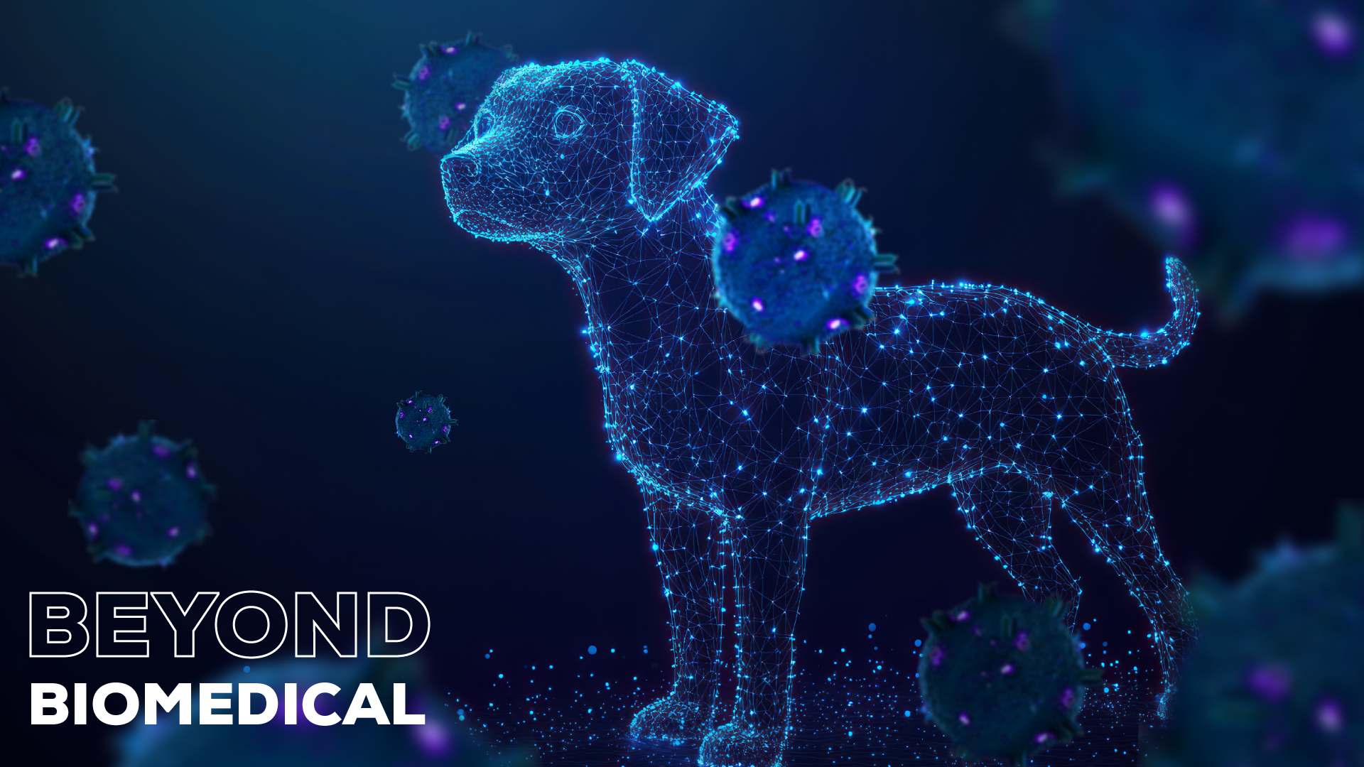


Could your pet’s urine one day reveal early signs of cancer, without a single invasive test?
Extracellular vesicles (EVs), such as exosomes and microvesicles, are gaining traction in human medicine applications, but did you know EVs hold promise in veterinary diagnostics and therapeutics, too?
From cancer diagnostics in dogs to kidney disease monitoring in cats, researchers are investigating the potential of EVs to transform health care for our furry friends.
And while EV research in veterinary medicine is still in its early days, momentum is building. As with human medicine, researchers face hurdles – like inconsistent methods for isolating, characterising, and storing EVs. On top of that, working with animals brings its own challenges, such as limited access to large study cohorts1. Still, new studies are emerging, and in this article, we explore how EVs might one day help improve care for animals large and small.
EVs as cancer biomarkers in dogs
We all want to make the best choices for our pets and that means we need to understand what’s wrong with them when they’re sick, and how their illness might progress. Mast cell tumours (MCTs) are one of the most common cancers affecting the skin in dogs, accounting for approximately 11% of skin tumours2. Breeds including Boxers, Labradors and Shar Pei are particularly prone to developing MCT2,3. Although they commonly affect the skin, MCTs are haematopoetic neoplasms that arise from mast cells, a type of white blood cell that resides in connective tissues and plays an important role in IgE-mediated immune response4. One challenge in the management of MCT is the variety of presentations, disease courses and clinical outcomes5, meaning both under- and overtreatment are possible.
Precision medicine – the tailoring of treatment to an individual – has been at the forefront of human medicine since the explosion of ‘omics technologies from the late 1990s onwards. A recent pilot study by Zamboni and colleagues aimed to develop a similar strategy for dogs with MCT. The study successfully used qEVoriginal 35 nm columns to isolate small EVs (sEVs) from blood samples from dogs with MCT, either with or without lymph node metastasis, and from healthy controls6. They found that levels of an sEV-associated microRNA, miR-21-5p, were significantly higher in dogs with nodal metastasis, which is correlated with worse prognosis and is a factor in deciding an appropriate treatment or management strategy3. Determining lymph node status is complex, as it is not always clear which lymph nodes are most likely to be affected, and the necessary mapping and biopsy are expensive and invasive procedures5. Zamboni et al. speculate that sEV-miR-21-5p could be developed as a prognostic biomarker to help overcome these barriers and determine the optimal treatment for each dog. They also highlight the follow up work that is needed to confirm this hypothesis6.
Canine carcinoma detection
Another key trend in human medicine in recent decades has been the development of liquid biopsies for cancer detection and monitoring. While this field started out developing blood sampling as an alternative to tissue biopsies, which are harder to obtain and pose greater risk to the patient, it has expanded to truly non-invasive biopsies, such as urine testing. Urothelial carcinoma (UC), also known as transitional cell carcinoma, is the commonest urinary tract cancer diagnosed in dogs and is particularly prevalent in breeds including Scottish Terriers and West Highland White Terriers7. Metastatic disease is frequent, but even local disease can be fatal due to urinary tract obstruction8. Given the severity of the prognosis, accurate diagnosis is essential and is currently achieved through invasive and specialised procedures such as catheterisation and biopsy7, making it a prime candidate for non-invasive liquid biopsy test development.
To try and simplify the detection of UC, Karttunen et al. used qEVsingle 70 nm columns to isolate EV-rich and protein-rich fractions from the urine of dogs with UC and compared them to those from dogs with urinary tract infections (UTIs) or healthy controls9. Their aim was to identify biomarkers that could be used in diagnosing UC via a non-invasive liquid biopsy sample, rather than cells collected by catheterisation. They used miRNA sequencing to screen 159 miRNAs for differential expression and followed up on the three strongest candidates - miR-182, miR-221 and miR-222. Of these, miR-222 performed best as a biomarker for UC, with an area under the curve (AUC) for cancer vs a mixed control group of healthy dogs & dogs with UTIs of 0.88 (95% CI 0.78-0.98). These results could pave the way for the development of not just diagnostic biomarkers for symptomatic cases but also provide candidates to investigate for screening test development that could be used for early detection in breeds predisposed to UC9.
Urinary EVs as a liquid biopsy in cats
Like dogs, cats also suffer from diseases where early, non-invasive detection could be beneficial. One key area of interest is kidney disease.
Chronic kidney disease (CKD) is a common condition in cats, occurring in 30-80% of cats over 15 years of age10,11. The underlying cause is frequently unknown, and disease course is variable10, hence there is a need for both biomarkers and therapeutic targets to improve monitoring, management options and, ultimately, outcomes. Urinary EVs are of huge interest as biomarkers of urinary tract physiology for a variety of conditions12 and feline CKD is another potential application.
Lawson and colleagues analysed urine EVs (uEVs) from cats with CKD, with either normal or high blood pressure—a common comorbidity—and healthy controls. They confirmed that urine concentration by ultrafiltration followed by size exclusion chromatography (SEC) using qEVsingle 70 nm columns is a suitable method for obtaining a good yield of EVs for proteomic analysis, an important first step in enabling future EV research. While the authors did not find any significant differences in uEV size, quantity or proteome between cats with CKD and controls, they did find several differentially expressed proteins between CKD-affected cats with and without hypertension. This raises further research questions, such as whether these changes are causative and whether they would also be found in cases of hypertension without CKD13.
Just the beginning of EVs in veterinary research?
The studies highlighted here show that EVs can be successfully isolated from animal biofluids and used for a variety of downstream testing to study disease states. EV research is being carried out on everything from fertility in horses14 to mastitis in cows15. Veterinary research is also expanding to therapeutic applications of EVs16. While there is still a long way to go, it seems likely that EVs will have a place in the future of veterinary medicine, just as they do in human medicine.


